What is Cervix?
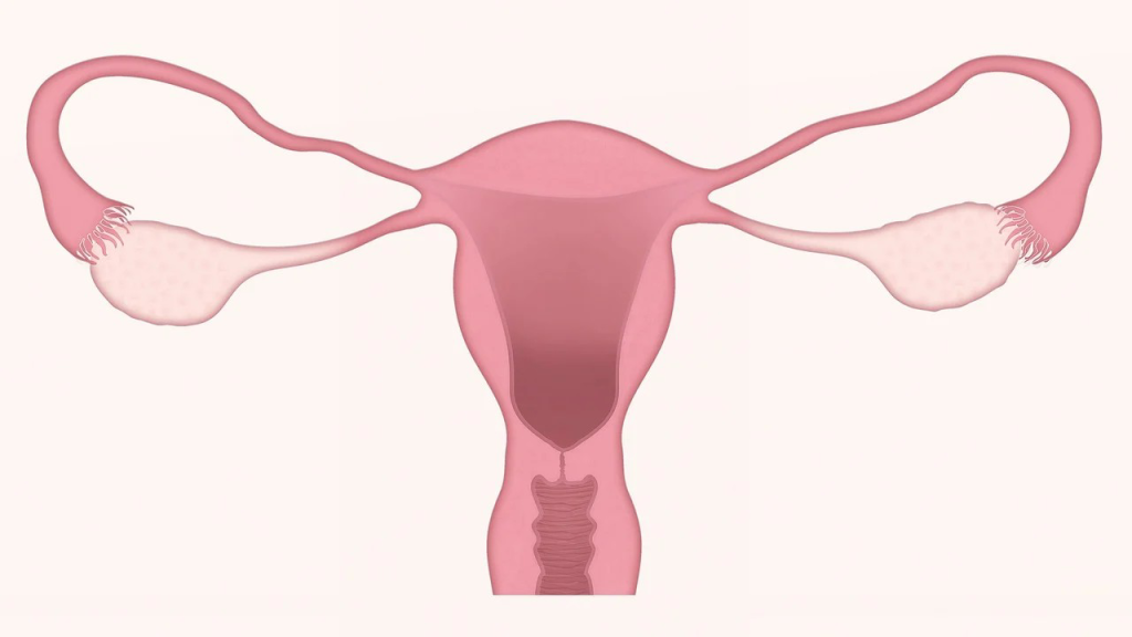
Cervix otherwise called uteri cervix or “neck of the uterus” refers to a cylindrically shaped fibromuscular structure that connects the uterus to the vagina. Strawberry cervix occurs there is cervical trichomoniasis characterized by bloody sightings over the vagina and cervix.
Position: It forms the lower one-thirds of the uterus. It is situated in the true pelvis and enters the vagina at right angles.
Shape: It is cylindrical in shape, while its cavity is spindle or cylindrical shaped and slightly fuse-form.
Size: It is approximately 2.5cm in length and 1.3cm thick.
Table of Contents
Gross structure
Macroscopically, the cervix is divided into:
The supra-vaginal part: This part lies above and outside the vagina. It’s also called ectocervix.
The infra-vaginal part: This is the portion lying with the vagina and is otherwise known as endocervix.
The transformation zone(TZ) is where there is overlap between the endocervix and ectocervix and remains the commonest site for cervical cancer.
The widest part of the cervix lies in the centre with a constriction above where it communicates with the body of the uterus known as the internal os and a constriction below where it communicates with the vagina called the external os.

Microscopic structure
The cervix unlike the body of uterus contains less muscular but more elastic tissues just like the body of the uterus it has three layers (within outwards)
- Endometrium
- Myometrium
- Perimetrium
Endometrium: This refers to Inner lining of ciliated epithelium containing racemose glands. It is arranged in folds giving a tree- like appearance termed “arbor vitae”
The myometrium: The cervical muscle fibres are arranged into two (2) fibres as follows:
- Inner layer of circular fibres which are thickly arranged and allow it to dilate during labour
- Outer layer of longitudinal fibres which extends from the body of the uterus. These fibres cause the cervix to shorten during labour.
Perimetrium: This covers that part of the cervix which lies anteriorly and posteriorly above the vagina i.e. supravaginal portion with the exception of that area lying in contact with the base of the bladder.
Blood, Nerve & Lymphatic supply
- Blood supply: It is through the uterine arteries.
- Venous Drainage:Through corresponding uterine veins
- Lymphatic Drainage: Into the internal iliac and sacral gland
- Nerve Supply: Sympathetic and parasympathetic from the Lee Frankenhausers plexus.
Anatomical surroundings
The cervix is surrounded by
- Anteriorly: Uterovesical pouch and the bladder.
- Posteriorly: Pouch of Douglas and the rectum.
- Laterally: Broad ligament and ureters
- Inferiorly: The vagina.
Functions
- It allows the passage of menstrual flow during menstruation.
- It helps to prevent infection entering the uterus by serving as a gateway to the uterus.
- The cervical mucus produced by the cervical glands changes its consistency during the menstrual cycle either to promote or prevent pregnancy.
- It Dilates and withdraws during labour to enable vaginal delivery of the baby and placenta
- Following delivery, it returns almost to its non-gravid state(involution).
Changes during pregnancy
During pregnancy, the cervical glands produce mucus which forms a plug of operculum that fills the cervical canal and helps to prevent infection of the genital tract.
Towards the end of the pregnancy,it feels very much softer and the internal os begins to dilate. This is known as ” cervical ripening”.
Changes during labour
When labour begins, the muscular fibre surrounding the internal os are drawn upwards by the retracted upper uterine segment and it is shortened as it arranges to form part of the lower uterine segment. This is called cervical effacement(taking up of the cervix).
Collectively, these physiological processes result in dilatation and subsequent delivery.
Changes during puerperium
Following delivery, it begins to close and return to its pregravid state. The internal and external os and canal between them must have reformed.
The external os is however, never completely closed but becomes a slit-like aperture which can admit a tip of finger. This is known as “multip’s os”. Hence, the cervix of a parous woman appears larger than that of a nulliparous woman.
Postnatal period examination usually carried out 6 weeks after delivery helps to review the status of the mother during the period of recuperation to detect any deviation such as subinvolution, postpartum infection, puerperal psychosis or mastitis.
Note that postmenopausal women have thinner cervix than that of those within reproductive age.
Cervical disorders
Healthy cervix is a key factor in maintaining a healthy pregnancy. However, sometimes, a disruption in its structure or function resulting disorders such as:
Cervicitis: Cervicitis refers to the cervical inflammation which is usually caused by infections such as chlamydia, gonorrhea and herpes simplex. Its clinic features are vaginal discharge and postcoital bleeding.
However, gonococcal and chlamydial cervicitis are often characterized with cervical oedema, mucopurulent discharge and cervical friability. While ulcerative lesions(multiple small vesicular lesions) point at Herpes simplex viral infection.
Treatment for infectious cervicitis involves administration of antibiotics for 7 days therapeutic regimen as well as sticking to safer sex practices.
Cervical incompetence: This is usually diagnosed during pregnancy when the cervix begins to dilate earlier, predisposing to premature labour and delivery. Its causes may include surgical procedures on the cervix such as dilatation and curettage(D&C). The incompetency is treated with cervical cerclage.
Cervical cancer/myomas: This occurs when there is cancerous growth on the cervix. It accounts for 6% of all leiomyomas which is often caused by infection of the human papillomavirus (HPV). The HPV also causes genital and cervical warts.
Its clinical manifestations are feeling of mechanical pressure, urinary urgency, dysuria, dyspareunia, urethral and ureteral obstruction, obstruction of the cervix, menorrhagia and dysmenorrhea.
Diagnosis of cervical cancer is through pelvic examination and ultrasonography. While its treatment includes chemotherapy, radiation and surgery.
Postcoital bleeding: This refers to bleeding after sexual intercourse and its causes may include benign or malignant etiology found on the cervix or other genital area such as cervical intraepithelial neoplasia (CIN) and invasive cancer, vaginal, or endometrial cancer.
If you are experiencing bleeding after sex, kindly meet a doctor (gynecologist) for a colposcopic examination and prompt attention.
Cervical Polyps/ Endocervical Polyps: These are abnormal small growths often painless and harmless found on the cervix which can result in vaginal bleeding.
The cervical polyps are diagnosed during pelvic examination and the patient may report postcoital, intermenstrual and/or postmenopausal bleeding. Where necessary, treatment involves simple surgery that can be performed in the office.
Cervical Ectropion
This is otherwise known as cervical ectopy or erosion and it refers to a condition where there is eversion of the endocervix exposing the columnar epithelium to the vaginal wall.
Adolescents and pregnant women including those taking estrogen-containing contraceptives are far more likely to have ectropion.
Its clinical features are vaginal discharge and postcoital bleeding(especially in pregnant women). Treatment is not needed unless there is excessive mucus discharge or troubling spotting. Cryotherapy or electrocautery is used for treating symptomatic ectropion.
Other cervical disorders are: Nabothian cysts (mucinous retention cysts or epithelial cysts) cervical dysplasia/cervical intraepithelial neoplasia (CIN), cockscomb cervix, cervical collar, and clear cell adenocarcinoma(CCA).
Tofort Recommendation
Cervix is a spindle-shaped organ that links the uterus to the vagina. It remains the key part of the female reproductive system. When placenta implants to the cervix, placenta praevia occurs, which is associated with increased risk of antepartum and and postpartum haemorrhages.
We therefore encourage every woman to take good care of herself by going for regular cervical cancer screening, treatment of underlying sexually transmitted infections and practicing safer sex.
By following these tips, you are far more likely to have a healthy cervix and live optimally. It’s our greatest desire to see you happy and healthy!