Cord prolapse: Causes, signs, and management

What is cord prolapse? Cord prolapse is an obstetrical emergency whereby the cord lies in the front of the presenting part with the membranes ruptured. It requires the expertise of an experienced midwife or obstetrician to save the baby’s life. During this period, the midwife should allay the woman’s anxiety and ensure that he or she provides woman-centered care devoid of disrespect and abuse. Proper information regarding what is happening should be provided to her and her support person or partner. Risk factors for umbilical cord prolapse Diagnosis A loop of cord may be visible below or beside the presenting part. It can be felt on vaginal examination. It may also be on the Os as in high head. Cord prolapse is diagnosed when membranes are ruptured.  General management of cord prolapse Immediate care Relieve pressure on the cord  by: Specific management of cord prolapse This depends on the stage of labour at which the diagnosis was made.  First stage: Fetus still alive – delivery not immenient or woman cannot deliver by vagina, Caesarean section is the treatment of choice. Second stage: Encourage mother to push with contractions. Perform episiotomy. If cephalic presentation, assist labour with vaccum or forceps. Community: Fetus is still alive – transfer to hospital immediately. Relieve pressure on cord by placing mother in left lateral position and buttocks elevated.  Inform consultant: prepare for emergency Caesarean section on arrival. Complications of cord prolapse Poorly managed umbilical cord prolapse is associated with dangers such as fetal hypoxia and death due to cord compression especially in premature and low birth weight babies. However, there are no specific ways of preventing cord prolapse. Pregnant women are usually advised to attend antenatal care regularly and utilize facility-based delivery where there are skilled and qualified birth attendants. Bottom line Umbilical cord prolapse is an emergency situation that needs obstetric interventions to save the life of the baby. When this condition occurs in the community, the midwife should carefully transfer to hospital where proper care will be provided. Again, midwife should avoid performing artificial rupture of membranes (ARM) in pregnant women diagnosed with polyhydramnios in order to minimize the risk of the cord prolapse due to the rushing of the liquor amnii. The midwife should also endeavour to provide respectful and evidence-based care to the woman as well as involve her in decision pertaining her care.

Cord prolapse: Causes, signs, and management Read More »

Cervix Anatomy: Description, Functions, Changes during Pregnancy & More

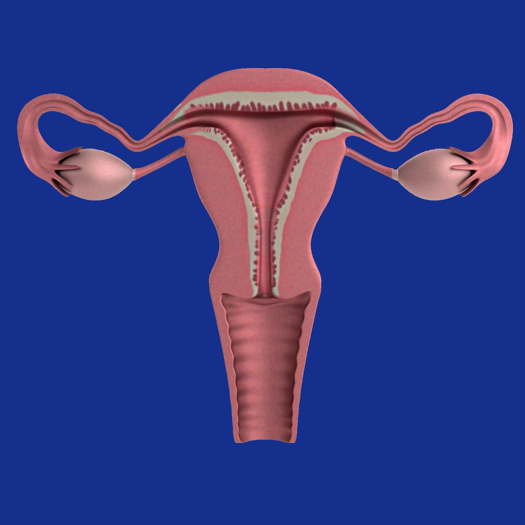
What is Cervix? Cervix otherwise called uteri cervix or “neck of the uterus” refers to a cylindrically shaped fibromuscular structure that connects the uterus to the vagina. Strawberry cervix occurs there is cervical trichomoniasis characterized by bloody sightings over the vagina and cervix.   Position: It forms the lower one-thirds  of the uterus. It is situated in the true pelvis and enters the vagina at right angles.  Shape: It is cylindrical in shape, while its cavity is spindle or cylindrical shaped and slightly fuse-form. Size: It is approximately 2.5cm in length and 1.3cm thick. Gross structure Macroscopically, the cervix is divided into:  The supra-vaginal part: This part  lies above and outside the vagina. It’s also called ectocervix.  The infra-vaginal part: This is the portion lying with the vagina and is otherwise known as endocervix.  The transformation zone(TZ) is where there is overlap  between the endocervix and ectocervix and remains the commonest site for cervical cancer. The widest part of the cervix lies in the centre with a constriction above where it communicates with the body of the uterus known as the internal os and a constriction below where it communicates with the vagina called the external os. Microscopic structure The cervix unlike the body of uterus contains less muscular but more elastic tissues just like the body of the uterus it has three layers (within outwards) Endometrium: This refers to Inner lining of ciliated epithelium containing racemose glands. It is arranged in folds giving a tree- like appearance termed “arbor vitae”   The myometrium: The cervical muscle fibres are arranged into two (2) fibres as follows:  Perimetrium: This covers that part of the cervix which lies anteriorly and posteriorly above the vagina i.e. supravaginal portion with the exception of that area lying in contact with the base of the bladder. Blood, Nerve & Lymphatic supply Anatomical surroundings The cervix is surrounded by    Functions Changes during pregnancy During pregnancy, the cervical glands produce mucus which forms a plug of operculum that fills the cervical canal and helps to prevent infection of the genital tract. Towards the end of the pregnancy,it feels very much softer and the internal os begins to dilate. This is known as ” cervical ripening”. Changes during labour When labour begins, the muscular fibre surrounding the internal os are drawn upwards by the retracted upper uterine segment and it is shortened as it arranges to form part of the lower uterine segment. This is called cervical effacement(taking up of the cervix).  Collectively, these physiological processes result in dilatation and subsequent delivery.  Changes during puerperium Following delivery, it begins to close and return to its pregravid state. The internal and external os and canal between them must have reformed.  The external os is however, never completely closed but becomes a slit-like aperture which can admit a tip of finger. This is known as “multip’s os”. Hence, the cervix of a parous woman appears larger than that of a nulliparous woman. Postnatal period examination usually carried out 6 weeks after delivery helps to review the status of the mother  during the period of recuperation to detect any deviation such as subinvolution, postpartum infection, puerperal psychosis or mastitis. Note that postmenopausal women have thinner cervix than that of those within reproductive age. Cervical disorders Healthy cervix is a key factor in maintaining a healthy pregnancy. However, sometimes, a disruption in its structure or function resulting disorders such as: Cervicitis: Cervicitis refers to the cervical inflammation which is usually caused by infections such as chlamydia, gonorrhea  and herpes simplex. Its clinic features are vaginal discharge and  postcoital bleeding.   However, gonococcal and chlamydial cervicitis are often characterized with cervical oedema, mucopurulent discharge and cervical friability.  While ulcerative lesions(multiple small vesicular lesions) point at Herpes simplex viral infection.  Treatment for  infectious cervicitis involves administration of antibiotics for 7 days therapeutic regimen as well as sticking to safer sex practices.  Cervical incompetence: This is usually diagnosed during pregnancy when the cervix begins to dilate earlier, predisposing to premature labour and delivery.  Its causes may include surgical procedures on the cervix such as dilatation and curettage(D&C).  The incompetency is treated with cervical cerclage.   Cervical cancer/myomas: This occurs when there is cancerous growth on the cervix. It accounts for 6% of all leiomyomas which is often caused by infection of  the human papillomavirus (HPV). The HPV also causes  genital and cervical warts. Its clinical manifestations are feeling of mechanical pressure, urinary urgency, dysuria, dyspareunia, urethral and ureteral obstruction, obstruction of the cervix, menorrhagia and dysmenorrhea.  Diagnosis of cervical cancer is through pelvic examination and ultrasonography. While its treatment includes chemotherapy, radiation and surgery. Postcoital bleeding: This refers to bleeding after sexual intercourse and its causes may include benign or malignant etiology found on the cervix or other genital area such as cervical intraepithelial neoplasia (CIN) and invasive cancer, vaginal, or endometrial cancer.  If you are experiencing bleeding after sex, kindly meet a doctor (gynecologist) for a colposcopic examination and prompt attention.  Cervical Polyps/ Endocervical Polyps: These are abnormal small growths often painless and  harmless found on the cervix which can result in vaginal bleeding. The cervical polyps are diagnosed during pelvic examination and the patient may report postcoital, intermenstrual and/or postmenopausal bleeding.  Where necessary, treatment involves simple surgery that can be performed in the office. Cervical Ectropion This is otherwise known as cervical ectopy or erosion and it refers to a condition where there is eversion of the endocervix exposing the  columnar epithelium to the vaginal wall. Adolescents and pregnant women including those taking estrogen-containing contraceptives are far more likely to have ectropion.  Its clinical features are vaginal discharge and postcoital bleeding(especially in pregnant women).  Treatment is not needed unless there is excessive mucus discharge or troubling spotting. Cryotherapy or electrocautery is used for treating symptomatic ectropion. Other cervical disorders are: Nabothian cysts (mucinous retention cysts or epithelial cysts) cervical dysplasia/cervical intraepithelial neoplasia (CIN), cockscomb cervix, cervical collar, and clear cell adenocarcinoma(CCA).  Tofort Recommendation Cervix is a spindle-shaped organ that links the uterus to the vagina. It remains

Cervix Anatomy: Description, Functions, Changes during Pregnancy & More Read More »

Fallopian tubes: Overview, Anatomy & Functions 

Description of Fallopian tubes The Fallopian tubes are muscular canals extending from the cornua of the uterus and opening into the peritoneal cavity near the ovaries.  It helps to move released or fertilized eggs to the uterus for implantation. Blocked fallopian tubes is one of the causes of infertility in women. Fallopian tubes are also called oviducts, uterine tubes or salpignx, which are conduits through which spermatozoa pass through to fertilize the ovum at the ampullary portion of the tube.  Each tube measures 10cm in length and 6mm in diameter and is enveloped in the upper poles of the broad ligament. The tubes have a lumen which communicate with the uterine cavity medially and opens into the peritoneal cavity laterally. Gross structure of the Fallopian tubes Each tube is described in 4 parts The Interstitial part:This is the narrowest part of the tube measuring 1.23cm in length and lying within the thickness of the uterine wall. It’s lumen measures 1 mm in diameter. The Isthmus: This is a narrow portion of the tube, extending for about 2.5cm laterally from the uterine walls. The Ampulla: This is much wider than the Isthmus, extending for about 5 cm from the Isthmus towards the side wall of the pelvis. Fertilization of the ovum takes place in the . The Infundibulum: This is the last of the tube which measures 2.5m & turns backwards and downwards. It is funnel shaped and composed of numerous finger- like processes/ projections called the fimbriae which surrounds the tubal orifice. One fimbra is attached to the ovary and is known as “the fimbria ovarica” and is the longest of all. Microscopic structure of Fallopian tubes Each  uterine tube has  three layers:  The Endometrial layer: This is an inner lining of mucous membrane thrown into complicated folds known as the plicae. The folds are designed to slow down the passage of the ovum to the uterus .  Note that the mucous membranes in the cervix are arranged in tree-like structure known as arbo vitae. The plicae are more developed in the ampullary part of the ovidicts. Many of the cells of the lining are made of cubical epithelium and are ciliated.  The non-ciliated cells are called goblet cells and  they secrete mucus into lumen of the tube shortly before menstruation. Beneath the inner lining is a layer of vascular connective tissue. The Muscular layer: The muscle coat consist of  two layers: (a) an inner circular layer which surrounds the mucous layer and (b) an outer layer of longitudinal smooth muscles.  The peritoneal layer: This is an outer covering of peritoneum which is absent along the inferior surface of the tube between the layers of broad ligament. Anatomic surroundings Blood, Nerve & Lymphatic supply to Oviducts  Functions of Fallopian tubes  Disorders of Oviducts

Fallopian tubes: Overview, Anatomy & Functions  Read More »

Uterus anatomy: Description, Functions & its physiology During Pregnancy

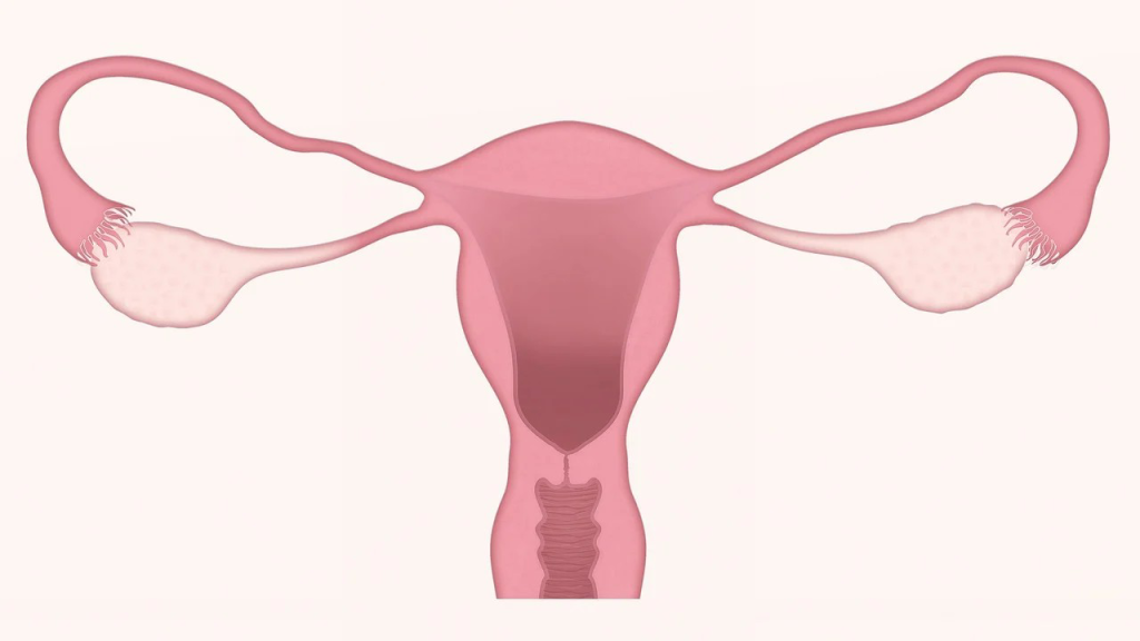
Description of Non-pregnant or non-gravid uterus Uterus is a pear-shaped, hollow muscular organ which receives the insertion of two oviducts into its upper and outer angle.  The uterus provides an environment for nourishment, growth and development of fetus during pregnancy till delivery. Position: The uterus is located within the true pelvis above the vagina behind the bladder and in front of rectum in an anteverted and anteflexed manner.  Size: Measures 7.5cm long, 5cm wide, 2.5cm depth and 1.25cm thick in each wall. The uterus weighs about 60g. Gross structure  The uterus is discribed in two parts which are the body and the cervix.  The body is the upper two-thirds of the uterus and this in turn consists of the following parts: (a) Fundus: this is the broad rounded upper portion above the insertion of the tubes.  (b) Cavity: it is the hollow ‘D’ shaped area which has its base above and apex below and measures 5cm. Due to the flatness of the uterus, the anterior and posterior walls are in apposition.  (c) Cornua(horn): This is the lateral angle of the uterine body where fallopian tubes insert.  (d) Isthmus: This is the constricted or narrowed part which joins the body to the neck. Isthmus measures 7mm in length and is selected immediately above the internal is. Its function is to expand during the 2nd trimester of pregnancy to form lower uterine segment. Cervix: This is the neck of the uterus. Cervix is spindled shaped portion of the uterus, which is described in two parts: the supravaginal  and the intravaginal portions. Macroscopic Structure  The walls of the uterus are mainly composed of plain muscle cells called the myometrium. The uterine cavity is lined by mucous membranes known as the endometrium and  the peritoneum that covers the body the body of the uterus is known as perimetrium. Endometrium: This is the inner linning of the mucous membrane. The coporal endometrium undergoes series of changes from puberty to menopause(i.e. its appearance varies with each day of menstrual cycle). During menstruation, it is shed as far as the basal layer.  The cervical endometrium are of poor quality and do not undego any changes. The cervical endometrium is thrown into folds called arbo vitae (fern-like appearance) Myometrium:  This is the plain muscular layer which forms the second middle coat of of the uterus. It forms ⅞th of the thickness of the uterine walls and its fibres are arranged in three patterns: The Perimetrium The outer part of peritoneum covers only the upper part of the uterus. It is reflected posteriorly to the rectum — forming the pouch of Douglas and also anteriorly to the bladder —forming the utero-vesical pouch.  Control of haemorrhage(action of living ligatures) During third stage of labour, the oblique fibres which are arranged in the figure of ‘8’ patterns contract rhythmically. When these fibres contract, they ligate the arteries that pass via them thereby arresting bleeding. In addition, the contractions of the oblique fibres result in occlusion of the blood vessels. When the vessels are ligated, the fibrins form at the cut- end of the vessels and clot forms which occlude the vessels — making the escape of blood to be controlled. That is haemostasis mechanism.  Anatomic relationships Anteriorly: bladder, utero-vesical pouch, coils of intestines Posteriorly: rectum and pouch of Douglas Superiorly: coils of intestines Inferiorly: vagina and the base of bladder Laterally: Fallopian tubes, ovaries and broad ligaments  Supports of the uterus  The uterus is maintained or held in its position by 6 pairs of ligaments and individually by the pelvic floor muscles.  The supporting ligaments are: Blood, Nerve & Lymphatic Supply to Uterus Uterine Functions Changes that occurs in the uterus during pregnancy  Position: By 12th weeks of pregnancy, the uterus rises out of the pelvis to become an abdominal organ.  It is more vertical than anteverted and anteflexed.  Shape: As the uterus is increasing in size, its shape becomes modified. At the onset of pregnancy, it’s pear-shaped.  But by the end of third month, it becomes globular. Between 12th and 36th weeks, it becomes ovoid as the fetus grows longer. By the end of the third month, the isthmus widens out to form the lower uterine segment. Size: As the pregnancy advances, the uterus with 60g weight and  7.5cm long, 5cm wide and 2.5cm thick becomes 30cm long, 23cm wide and 20cm thick and weighs 900-1000g. The Isthmus increases from 7mm to 25mm in length. Changes in myoctyes(uterine muscle cells) The uterine muscle growth during pregnancy occurs in two ways which are: Hypertrophy: The actual muscle cells enlarge, increasing ten times in length and five times in width.  Hyperplasia: New muscle cells appear and grow along side of the pre-existing muscle cells.  Other changes occurring during pregnancy are: Physiology of uterus during pregnancy The uterus does not only enlarge remarkably to accommodate the growing fetus but also relaxes in order not to expel the fetus before term(uterus is intrinsically contractile). The growth and relaxation of uterine muscles are due to the hormones, oestrogen and progesterone respectively, both from corpus luteum and later from the placenta.  The uterus, however, is not completely relaxed. This is  because from 8th weeks onwards, periodic waves of contractions pass via the uterus. This lasts only for one minute and reoccur at  5-10 minutes intervals.  They are painless and women are unaware of their presence. These uterine contractions are known as “Braxton Hicks contractions”, “false labour” or “practice contraction” because they are simply preparing uterus for the role it would play during labour.  Towards the end of pregnancy, the contractions become stronger and more frequent and are responsible for taking up of the cervix(effacement) during the last month of pregnancy.  They increase during labour and become true labour contractions, and the woman then experiences discomfort or pain.  The bottom line  The uterus otherwise known as womb, is a pear-shaped muscular organ of the female reproductive system which is suitated  between the bladder anteriorly and the rectum posteriorly. It houses and nourishes fertilized egg (zygote) till

Uterus anatomy: Description, Functions & its physiology During Pregnancy Read More »

What are the birthing positions in labour?

Birthing positions Types of birthing positions RECUMBENT POSITION (LITHOTOMY) Women can choose a variety of positions for birth. Use of non lithotomy positions were natural ways of birth as old as origin of man. In modern times about two centuries ago, the use of lithotomy became the norm because it is easier to apply new technology. It is now advocated that women can choose any comfortable position ranging from standing, sitting, squatty, hand and knees or side lying (left lateral) positions This is more convenient for health workers (midwives and others) to enhance maintenance of asepsis, assessment of fetal heart rate(FHR) and giving of episiotomy and repair. Nevertheless, it has following disadvantages to the comfort of the woman and the fetus LEFT LATERAL POSITION This is a common position some women and birth attendants always use. The woman lies on her left side with the left leg extended and her right knee drawn against her abdomen or flexed by her side. Although frequency of contraction may decrease in this position, the intensity increases leading to greater efficiency Advantages Disadvantages SQUATTING POSITION Squatting primarily common choice for some women because it favours gravity and the abdominal wall is relaxed.  Other advantages: Disadvantages SEMI-FOWLER’S (HALF SITTING) POSITION Most birth attendants advocate for this position. It is halfway between sitting and recumbent positions Advantages SITTING POSITION This can be used where there is availability of delivery chairs. This method can be traced back to ancient Egypt and was widely used in Greek and Roman civilizations.  In the early 19th century use of delivery chairs was discouraged and its use diminished on hygienic grounds due to increase in puerperal fever. A supported sitting position can be achieved by use of a support person in a delivery bed (couch). Advantages  Disadvantage It carries a potential for increased blood loss. HANDS AND KNEES POSITION Advantages Disadvantages Tofort’s Recommendation on choice of birthing positions 

What are the birthing positions in labour? Read More »

Second stage of labour: Description,  Physiology & Management

Description of Second stage of labour Second stage of labour starts from fully cervical dilatation to the delivery of the baby. It is also divided into two phases Latent phase of the second stage The cervix is fully dilated but the presenting part is still very high, it has not reach the pelvic outlet Active phase of the second stage The presenting part of the fetus reaches the pelvic outlet and with the presenting part on the perineum, this will stimulate the nerve at the perineum causing expulsive Uterine contraction or urge to bear down during contraction and is also known as Ferguson’s reflex. Physiology of second stage of labour Uterine Action Soft Tissues Displacement Signs and symptoms of second stage The following signs are indicative of second stage of labour. They are classified under presumptive and confirmatory evidence PRESUMPTIVE SIGNS The following signs CONFIRMATORY EVIDENCE Vaginal examination must be taken in the presence of these signs to confirm full dilatation and ensure that the woman is not pushing too early. It also help to time second stage of labour Management of second stage of labour Assessment during second stage of labour Assessment of the woman and the fetus is continuous during second stage of labour. There are four determinants of outcome of second stage and they must be carefully monitored. They are as follows: UTERINE CONDITIONS The midwife should observe the strength duration and frequency of uterine contractions during second stage. It is usually longer and stronger during the first stage of labour with longer resting phase. The position the mother adopt for delivery usually influences the contractions. The midwife observes contractions through maternal response and abdominal palpation. DESCENT, ROTATION AND FLEXION In primigravida, descent may be slow during latent phase of second stage and accelerates during the active phase.  In multigravida,  the descent occurs more rapidly if descent is not progressive despite good uterine contractions and good maternal pushing on abdominal palpation, vaginal examination should be carried out to assess the station of the presenting part, whether or not internal rotation has taken place and rule out excessive caput succedaneum.  The labour is likely to progress well if the occiput has rotated anteriorly ( well flexed head) and there is no excessive caput, the midwife should continue.  Where there is no good rotation and flexion, poor contractions change position, give her nourishing fluid for hydration. Consult more experienced midwife. If fetal or maternal condition is compromised obstetrician should be involved in the management FETAL CONDITION Thick fresh meconium stained liquor when the membrane ruptures indicate fetal compromise (distress) and obstetrician should be called immediately. Fetal heart rate is checked after every contraction in the second stage of labour using pinnard stethoscope or sonicaed. Fetal distress is suspected if the following are observed If this is observed for the first time, change the mothers position because this may be as a result of head compressing the cord.  However if it persists, give episiotomy if delivery is imminent to expedite delivery. Seek an experienced midwife to expediate delivery with use of vacuum extractor MATERNAL CONDITION The midwife observes both psychological and physiologic parameters of the woman during second stage of labour. The woman’s ability to cope emotionally is assessed.  During the second stage there is increased apprehension or irritability. Some women may cry or make a lot of expression. These helps the midwife assesses woman’s coping ability. Also assess physical well being by checking maternal pulse rate ½ hourly and blood pressure. Conduct of normal labour Principles of Delivery These includes Preparation for the birth Before delivery of the baby the midwife should prepare for the birth Promoting maternal comfort in second stage of labour Most mothers feel very hot and exerted during the second stage, they sweat profusely due to pushing, a cool towel should be applied to the face, neck and body and these provide soothing.  The lips and tongue are dried and cracked and saliva sticky. Sips of water or other fluids will be very smoothing.  Ensure that the bladder is empty to avoid trauma to the bladder (pressure of the presenting part on the full bladder) Delivery of the baby The midwife should help position the woman (adopt the position she chooses) Birth of the shoulder Final notes on second stage of labour

Second stage of labour: Description,  Physiology & Management Read More »

First stage of labour: Description,  Physiology & Management

Description of first stage of labour The progress of labour has been divided into stages and phases, labour is subdivided into four stages they are as follows:  FIRST STAGE OF LABOUR This begins with the onset of true labour and ends when the cervix is completely dilated at 10cm.  This stage is subdivided into three phases as follows:  Latent phase This begins with onset of irregular Uterine contraction, the cervix begins to dilate and efface little or with no fetal descent. The cervix dilates from 0 – 4 cm, it lasts for 6 – 8 hours in primip gravida and shortens in multigravida.  The cervical canal shortened from 2 – 5 cm to less than 0.5cm. Uterine contraction becomes established during this phase and increases in frequency, duration and intensity Active phase This is characterized by rapid cervical dilatation, initially when two phases of the first stage were recognized. Active phase begins from 4 cm dilation to 10 cm. The cervix dilated 1 cm per hour, fetus descent is progressive, contraction occurs at a frequency of 2-5 times in 10 minutes.  Moderate to strong uterine contractions lasting for 2- 40 seconds progressing to greater than 40 – 60 second s. The woman’s anxiety increases as contraction and pain increases, she may adopt various coping mechanisms Transition phase This is from 8 cm dilation to 10 cm, this is the last part of the first stage and the woman demonstrates significant anxiety.  The contractions becomes more intense and forcefully, the woman becomes restless and frequency changing positions, the contractions are strong about 3 – 5 times in10 minutes lasting for 60- 90 seconds as dilatation approaches 10 cm, there is an increase in rectal pressure and uncontrollable urge to bear down, increase in bloody show, spontaneous rupture of membrane, if membrane has not been ruptured already.  There is a sensation of great pressure at the pick of contraction that it seems that her abdomen will burst open. Physiology of the first stage of labour The events of first stage of labour are divided into two major categories for better understanding, and they include: The duration of labour This varies depending on numerous factors such as parity, birth interval, psychological state, presentation and position of the fetus, maternal pelvis shape and size, and the nature of Uterine contraction. First stage of labour takes the greater part of the labour, especially the latent phase.  Active phase of labour is expected to be completed between 6 – 12 hours, labour last longer in primip gravida woman than multiparous woman, duration of labour should not last longer than 18 hours Note: World Health Organization (WHO) recommended management of the active phase of labour with pantographs, this will help manage the progress of labour and action to be taken if labour is prolonged. Uterine Action Series of event occurs in the Uterus during labour such as Management of first stage of labour

First stage of labour: Description,  Physiology & Management Read More »

Types of pelvis:Gynaecoid,  Android, Anthropoid, Platypelloid & their effects on labour

Type of pelvis and their effects on labour The shape and structure of pelvis varies from one woman to another, ut ideal normal pelvis favours vaginal delivery. There are four main types of pelvis are gynaecoid, android, anthropoid and platypelloid. Table of contents GYNAECOID PELVIS This is another name for the female pelvis designed for child bearing. It is found in about 50% of women whose hips are broader than their shoulders with a height over 1.5m and shoe size of about 4 and above.  Characteristics Effects of gnaecoid pelvis on labour The fetal head is always engaged in transverse diameter of the brim in an anterior position, this causes the mechanism of labour normal, therefore the gynaecoid pelvis is the best for child bearing. ANDROID PELVIS This is the male type of pelvis, possessing about 20% of women. The bones are heavier than the gynaecoid pelvis. Characteristics Effects of Android pelvis on labour The available space in this type of pelvis is restricted by the triangular shape of the brim, the head, therefore may engaged transversely in an occipito posterior position, so this type of pelvis favours: ANTHROPOID PELVIS This is an APE type of pelvis possessing about 24–25% of women who have very tall long legs with narrow shoulders. The bones are quite heavy, this type of pelvis is common in women in South Africa. Characteristics Effects of Anthropoid pelvis on labour PLATYPELLOID PELVIS This is a flat pelvis possessing about 5% of women Characteristics Effects of Platypelloid pelvis on labour

Types of pelvis:Gynaecoid,  Android, Anthropoid, Platypelloid & their effects on labour Read More »

Bony Pelvis: Description, Structure & Functions

General description of bony pelvis The bony pelvis is a strong bony ring linked to a tilted basin which protects and contains the female reproductive organs. The following are found within the pelvis:  It is through these route that a baby passes through during delivery. Structure of the pelvis The bony pelvis is made up of 4 bones these are: INNOMINATE BONES These forms the anterior and lateral walls of the pelvis. Each innominate bones consists of: ILIUM This is the largest flat plate of bone above and part of the acetabulum below. Characteristics Anteriorly: the iliac crest ends in the anterior superior iliac spine Posteriorly: it ends in the posterior superior iliac spine. THE  ISCHIUM This is the lowest of the constituent bone of the innominate bones. The shape is C curved. Characteristics THE PUBIS This is the smallest of the innominate bones and it is the only one that articulate with it’s fellow on the opposite side. Characteristics THE  SACRUM Is situated at the posterior part of the pelvis, therefore it forms the posterior wall of the pelvis. It is waged shaped Characteristics THE  COCCYX This is a small bone which consists of 4 fused coccygeal vertebrae. It is trianglar in shape with it base at the upper most Characteristics PELVIC  JOINT There are 4 in number  The sacroilliac  Joint The joint is formed at the articulates of the sacrum with the ilium in normal condition, very slight movement occurs at this joint. But during cyesis( pregnancy) and labour. This movement increases in range when the ligament becomes softened under the influence of the hormone relaxing The symphysis pubis Joint This joint is formed at the junction of the two pubic bones, which are United by a pad of cartilage. This joint widely appreciably during the later month of pregnancy and the degree of movement permit may give rise to pain on walking The sacrococcygeal Joint This joint is formed where the base of coccyx articulates with the tip of the sacrum and allows the coccyx to bend backwards during the actual birth of the head of the baby PELVIC  LIGAMENTS There are 6 pelvic ligaments which are of important to midwife. Supporting Ligament This is the strongest ligament in the whole body, they binds with the sacrum and ilium at the Sacro Illiac Joint Sacro Tuberous Ligament It is a strong ligament passing from the posterior superior iliac spine and the lateral border of the sacrum and the coccyx to the ischial tuberosity. It bridges across the greater and lesser sciatic notches. Note: the Sacrotuberous and spineous ligament forms the posterior wall of the pelvic outlet. Sacrospineous Ligament This pass from the sides of the sacrum and coccyx across the greater sciatic notch to the ischia spines. It is a strong ligament and lies in front of the Sacro tuberous ligament.  Inguinal Ligament This ligament runs from the anterior superior iliac spine to the pubic tubercle. Lucuna Ligament This is a small ligament which occupies the angle under the inner end of the inguinal ligament. Obturator Membrane This ligament closes the obturator foramen with the exception of small area in its upper part which transmit the obturator vessels and nerves and also contains the obturator lymphatic glands. THE REGIONS OF THE PELVIS Obstetrically the bony pelvis is divided into two as follows: The False Pelvis This is the part above the brim which consists mainly the flared out iliac bones. It has little obstetric importance. The True Pelvis This is the curved bony canal through which the fetus most pass during birth. It consists of the brim, the cavity and the outlet. . THE AREAS OF THE PELVIC MEASUREMENT (DIAMETERS) THE PELVIC BRIM This is the first part of the Pelvic gland, it is rounded accept where sacral promontory encroaches into it. The promontory and the wings of the sacrum form its posterior border (alae of the sacrum). The iliac bone forms it’s lateral bounder while the pubic bone forms it’s anterior bounder. Obstetrically, the boundaries of the brim also known as its landmarks, are very important to midwife and these includes:  THE  CAVITY This is circular in shape and it’s anterior walls form by the posterior pubic bones and the symphysis pubis. It measures 4cm, the posterior walls measures 12cm and is formed by the curve of the sacrum. Laterally are the greater sciatic notch,body of the sacrum and obturator foramen covered by the obturator internus muscles. THE  OUTLET This is described into two parts The Anatomical outlet The Obstetrical outlet The obstetric outlet is of greater important to midwife because it is the areas in which the fetus must negotiate as passes through the birth canal rather than the wide anatomical outlet. DIAMETERS OF THE PELVIS A diameter is a distance between two points. The study of diameter is of greater important to midwives for the skillful management of labour. DIAMETERS OF THE BRIM Four diameters are measured and they includes:  the sacrocondyloid is the smallest diameter of the brim and it is improtance is concerned with posterior position of the occiput when the pariental eminance of the fetal head may become trapped. DIAMETERS OF THE CAVITY There are 3 diameters of the cavity DIAMETERS OF THE OUTLET 3 diameters are obtained at the outlet and these includes:  TYPES OF CONJUGATE THE PLANES OF THE PELVIS & THEIR ANGLES OF INCLINATION The planes of the pelvis are imaginary flats surface drawn at various levels of the pelvis canal that is:  When a person is standing upright the plan makes an angle of 60 degree to the floor. Because the Pelvis is curved canal. The angles of the brim, cavity and outlet must be different too. Therefore the angle of the plane of the Pelvic brim to the floor is 60 degree. The angle of the plane of the cavity is 30 degree while that of the outlet to the floor is 15 degree. AXIS OF THE PELVIC CANAL This is an imaginary line drawn

Bony Pelvis: Description, Structure & Functions Read More »

Anatomy of Ovaries: Description, Function & Physiology of Ovulation

Description of Ovaries The Ovaries are the female glands or organs that lie within the peritoneal cavity in a small depression of the posterior wall of the broad ligament. Location: They are situated at the fimbrated end of the Fallopian tube at about the level of the pelvic brim. Shape: They are small almond like organs dull white in colour and with a corrugated surface Size: They measure 3 cm in length 2 cm  in breath and 1 cm in thickness. They weigh about 6 gram Gross structure This varies with the age of the woman. From birth to puberty, the organs are smooth dull white and rather solid in consistency. During the menstrual phase ie from puberty to menopause, the organs are longer and rather on the surface, and in more like a walnut than an almond.  During the post menopausal phase, the ovaries beco smaller and shruken and are covered with scar tissues, where month after month the graffian follicle have ruptured. Microscopic structure The ovaries are composed of the cortex and a medulla surrounded by a germinal epithelium. Changes begin in some of the primordial follicles right from the Uterine life and persist throughout infancy and early girlhood. These changes consist of increase in the number of calls around the ovum and is followed by the appearance of fluid between the cells known liquour folliculi and the primordial follicle is now known as the “graafian follicle” The tunica albuginea is the outer part of the corvex formed by a sense fimbrous coat. The germinal epithelium consists of a layer of low cubical cells which covers the tunica albuginea of the cortex and is continuous with the mesovarian. Therefore the germinal epithelium is the modified form of the peritoneum.   Ovarian Attachments Relations of the ovaries Blood supply: by the ovarian and Uterine arteries Venous Drainage: by corresponding veins Nerve Supply: by ovarian plexus Lymphatic Drainage: into lumbar glands. Functions of ovaries Hormonal secretion of the Ovaries The graffian follicles and corpora lutea are not only under the hormonal influence of pituitary gonadotropins, but also secrete hormones on their own account. During the development of the graffian follicle, oestradiol is secreted by the cells of zona granulosa and the theca internal and stored in liquour folliculi. After ovulation it is secreted in lesser amount by the corpus luteum and is also formed in the cortex of the supra – renal glands. It is metabolized in the liver and excreted mainly in the urine in form of oestriol and oestreone and some 8 other related compounds.  These substances are collectively called oestrogen and are responsible for the secondary sex characteristic of puberty in young girls and some changes that occur during the menstrual cycle and pregnancy. Effects of oestrogen Major effects of oestrogens are: Progesterone —Key hormone of ovaries The corpus luteum under the influence of LH produce another hormone known as progesterone and a small quantity of oestrogen. The changes produced in the body by progesterone are mainly manifested in large quantities. Apart from pregnancy, it’s main function is endometrial changes as part of the menstrual cycle. Effects of progesterone During pregnancy it is responsible for:  The Ovarian Cycle This is described in three parts as follows:  Development of graffian follicle During fetal life the cortex of the ovary forms thousands of primitive eggs cells or primordial follicles which are present when the child is born. Every month from age of puberty onwards through child bearing life several follicles begins to develop as a rule, only one becomes fully matured and rises to the surface of the ovary. The one that matured is known as the graffian follicle (named after Dr.Von Graff).  Structure of a mature graafian follicle A mature graafian follicle is about 8-12mm.  Mechanism of ovulation Ovulation means ripening and discharging of an ovum from a graffian follicle into the peritoneal cavity. The reproductive age of a woman begins with menarche (appearance of first menses) to menopause (cessation of menstruation) at about 45-50 years. During this period, one graffian follicle out grows it’s fellow and enlarges in size . It then protrudes to the surface of the ovary with the discus and the ovum. As the follicle enlao it stretches the ovarian capsule (covering) until it becomes so thin and breaks finally. The follicle thus ruptures and the liquor folliculi, Discus and ovum are cast out into the peritoneal cavity. This process is known as ovulation. Ovulation takes place each month in alternate Ovaries so that each individual ovary ovulates at 2 monthly intervals. The corpus luteum After ovulation the walls of the graffian follicle collapses and become wrinkled at the same time some of the cells grow and develop yellowish coloured bodies. This yellowish wrinkled structure is known as corpus luteum, which under the influence of LH produce by the anterior pituitary produce progesterone which thickens the endometrial wall. It remins active for 14 days but if after that period fertilization does not take place, it does ,shrinks and become corpus albicans.  This leaves a whitish scar on the surface of the ovary. On the other hand, if fertilization takes place corpus luteum persist for 12 weeks until placenta is formed to take over the hormonal function of the corpus luteum. Hormonal control of ovulation The growth and ripening of the ovarian follicle are under the hormonal control- Follicular stimulating hormone (FSH) from the anterior pituitary gland. FSH is carried in the bloodstream from the pituitary to the ovary where it causes the follicle to grow and enlarge and at the same time stimulate the cells of th zona granulosa and the theca internal to produce oestradiol. When the amount oestradiol reaches a certain level, the production of FSH is stopped and LH from anterior pituitary gland is produced. When the FSH is falling, LH is rising and at certain ratio , this cause the follicle to rupture and ovulation occurs. After ovulation, the development and growth of corpus luteum occurs due to action of

Anatomy of Ovaries: Description, Function & Physiology of Ovulation Read More »